The composition of cell tissue in a colorectal polyp, as seen by NBI, can be divided into three types called NICE classification:
NICE 1: The polyp’s cell tissue still has a normal composition. In this case, the doctor will order follow-up checks to monitor the development of the polyp. However, he/she will not need to remove the polyp because the chances of it being cancerous are still very slim.
NICE 2: The composition of the cell tissue looks similar to a net, which indicates that the cells could eventually become cancerous. The doctor will therefore remove the polyp immediately using a coil or a special type of electric knife.
NICE 3: The cell tissue appears wild and disorderly, much like cancer cells. In this case, the doctor may not remove the whole polyp using a colonoscope, but may use tissue acquisition instead. The patient will need a consultation with an oncologist to assess which method of removing the polyp is most suitable.
The use of the NBI technique gives a more precise diagnosis. It also makes it easier for the doctor to devise a treatment plan. NBI can identify even the smallest polyps, as little as 5mm in diameter. This, thus, allows the doctor to calculate as accurately as possible whether the polyps pose a risk to the rest of the body. With a white-light colonoscopy, however, such small polyps might go unnoticed.
Still, the effectiveness of any colonoscopy using NBI largely depends on the expertise of the doctor and his/her mastery of the technique. It depends specifically on his/her adeptness at reading the results of the epithelium screening. Moreover, since there are only a small number of doctors who are experts in this technique, hospitals often overlook NBI as a screening option. This should not be the case, however. Infact, NBI offers a safe alternative to chromoendoscopy, as doctors just need to inject the patient with a dye or stain.

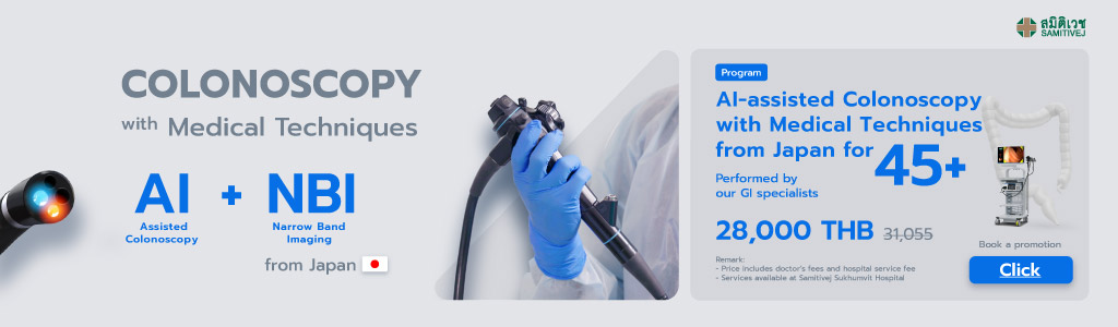
A Team of Expert Doctors and AI Technology from Japan
Samitivej Hospital, located in Bangkok, Thailand, has partnered with Sano Hospital in Japan, a facility world-renowned for its expertise in the removal of large polyps in the colon and early detection of gastric cancer using gastrointestinal endoscopy techniques. Since 2015, doctors from Samitivej’s Liver and Digestive Institute have traveled to Japan to train in colonoscopy and surgery, while doctors from SANO Hospital have come to Thailand to share their knowledge and conduct case studies on Samitivej patients.
At present, Samitivej is using AI technology in colonoscopy, along with NBI techniques from Japan that help double the accuracy rate in diagnosis. This collaboration and technological advancement have led to significant achievements by Samitivej in the field of gastroenterology, notably in the early detection and prevention of colon cancer:
- 13,595 cases of colon cancer prevented using AI
- Colon cancer incidence reduced by 60%
- Colon cancer fatalities reduced by 60 - 70%
- EndoBRAIN-EYE detects 1.5 times more polyps
- NBI techniques from Japan double diagnostic accuracy
- GI doctors at Samitivej Sukhumvit achieve a 55% average adenoma detection rate, compared with the 25% industry average have led to significant achievements by Samitivej in the field of gastroenterology, the American Society for Gastrointestinal Endoscopy (ASGE)
- The cost of cancer treatment reduced 20-fold