Breast Cancer: Screening and Prevention
HIGHLIGHTS:
- Breast cancer is more commonly found in younger Asian women than in Westerners, with 40-60-year-olds being particularly at risk.
- Undergoing a Digital Mammogram can reduce the chances of breast cancer mortality by up to 30% for females over the age of 40 years.
- Irregularities in the BRCA gene, which can be passed on from parents to their offspring, increase the risk of females developing breast cancer by almost 90% and for ovarian cancer by up to 50%.
Prevention against breast cancer depends mostly on early detection. Therefore, it is essential for every woman to know the different methods of screening and getting themselves checked up to stay ahead of the curve. Here is a guideline to screening and prevention which can benefit all women:
| Screening Method | Frequency | Who needs it? |
| From the age of 20 | ||
| Breast Self- Examination | On a monthly basis | All women from the age of 20 |
| From the age of 40 | ||
| Mammogram and Ultrasound | One or two times per year | All women from the age of forty, but especially women who:– Had children after the age of thirty– Had early menstruation (before the age of 12) – Had late menopause (after the age of 65) – Is childless – Has taken hormones or birth control pills over a long period of time -Has undergone radiation treatment for breast cancer – Women younger than 40 years old who have detected an abnormality during breast self-examination or those who are at risk for breast cancer, such as those with history of breast cancer in their family. |
| Under the age of 40 | ||
| Ultrasound | One or two times per year |
All women under the age of forty, but especially women who: – Have dense breast tissue (more common among Asian women than westerners) – Have encountered problems during a doctor’s examination or during a mammogram – Pregnant women who wants to avoid radiation |
| From the age of 30 | ||
| BRCA Gene testing | – | Women who have a history of breast cancer in their family |
Breast Self-Examination
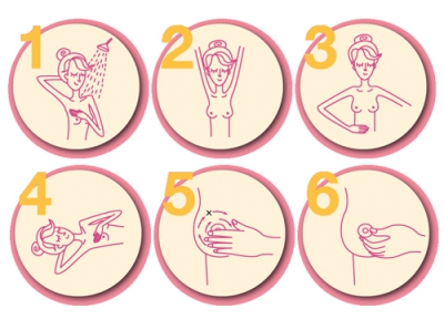
Start breast self-examination from the age of 20 on a monthly basis after your period ends, as during this time your breasts are least likely to be swollen. The method suggests that the woman stands in front of a mirror and inspects her breasts for abnormalities in several positions: with the arms hanging next to the body, arms held over head, and hands on the hips.
Things to observe when you are in front of the mirror
Observe the shape, size, skin texture and color of your breasts. If you notice any of the following abnormalities, consult your doctor immediately:
- Visible Lump
- Swelling, redness and warmth
- Changes in the size or shape of your breasts
- Dimpling or retracted nipples (if this is not the case previously)
- The skin on the breasts has the appearance of orange peel
- Rashes, scaling or itching around the nipple
- Abnormal discharge from the nipple such as blood-like fluid, or pus-like fluid
- The skin on the breast or nipples pulled inward

Breast self-examination palpation technique
Breast self-examination palpation technique uses the pads of your three fingers: index finger, middle finger and ring finger. The hand on the other side of the breast being examined is used to apply a circular motion, dragging the fingers around the breast without lifting them from its surface. The palpation needs to cover the entire breast from the nipple towards the tail and around the armpit. The palpation can take several forms of movements, such as circular spiral or zig zag (up and down motion) or radial pattern similar to the radius of the sun.
After one breast has been examined, perform the same on the other breast. If you feel a lump or if you are unsure it is a lump, do not hesitate to see your physician.
Digital Mammogram
This method takes x-ray of the breasts to assist the doctor in the diagnosis of breast cancer. The x-ray will show details of the abnormalities including any very tiny growth that cannot be detected by palpation of the breast, in particular in elderly women.
Studies show that the mammogram is an effective method in detecting early stage of breast cancer. Therefore women between 40-50 years old, who are in the high risk group for breast cancer, should get the examination every 1-2 years. Women who are over 50 years old are in the higher risk group and they should get the examination every year. During the x-ray process, the breast is compressed firmly against the film in order to get the clearest picture, so you will feel some soreness in the breast. Mammogram can detect growths which cannot be detected by palpating the breast, however there may be only 10% of the lumps that you can feel by palpating which cannot be seen in the x-ray
Benefits of Digital Mammography
- Less radiation from the x-ray process
- Easier detection; abnormal lumps can be identified more clearly and precisely, even ones that are only 0.1 – 1 cm. in size.
- If the images are not clear enough, the film’s resolution can be adjusted without the patient having to undergo the mammogram again
- It is a minimally invasive procedure that is safe for your breasts.
How to prepare for a mammogram?
- Do not apply powder, lotion or perfume to your armpits, breasts or chest area. This will affect the results of the mammogram
- Always bring in the films on your old mammogram so that the doctor can compare them to your new results
- Do not undergo a mammogram if you feel any strain or tension in your breasts. The ideal time to get a mammogram is 7-14 days after your menstrual period. During this time, the level of hormones in your body is decreased, resulting in less tension in your breasts and therefore, less pain during the mammogram
- There is no need to abstain from food or water
- If you have had breast surgery in the past, inform your doctor
- Wear a two-piece outfit rather than one. Do not wear earrings or necklaces.
Ultrasound
Today, combining mammogram with ultrasound is common for examining and diagnosing breast cancer. Ultrasound can diagnose if the lump is a fluid filled sac or muscle growth. It is suitable for patients with small breasts or patients during their childbearing period (with heavy breasts or breasts full of mammary glands) and patients with augmented breasts. It is also suitable for pregnant women who want to avoid x-ray radiation.
The use of Ultrasound in conjunction with Mammogram to detect breast cancer will result in a much more accurate diagnosis, especially for Asian women with dense breasts. Ultrasound can detect an abnormal lump that is as small as 2-3 millimeters.
BRCA and Breast Cancer
One of the most common causes of breast cancer is the genetic mutation found in BRCA, which can be divided into BRCA1 and BRCA2. The BRCA mutation is a hereditary condition that can be passed on by both the father and the mother to their offspring. The BRCA mutations in women increase a life time risk of 90% for breast cancer and 50% for ovarian cancer. Those who discover this hereditary condition can undergo breast or ovarian surgery to prevent cancer.
Learn more about BRCA and the necessity of genetic analysis here.
References
- The New York Times – Calls for More Genetic Testing for Cancer. Available from http://www.nytimes.com/2014/09/09/health/lasker-award-winner-calls-for-more-genetic-testing-in-cancers.html . Accessed on September 23, 2015.
Related
articles


